While in the hospital to have Heidi's teeth cleaned, we found two little bumps on her back legs that turned out to be mast cell tumors. This account is about how we found them, what they are, and how they were removed.
In a previous post, SMILE - Pre-anesthetic Physical and Pre-Medication, I mentioned how the licensed veterinary technician had a difficult time placing Heidi's IV catheter. Sometimes this can be because the animal is not cooperating - they tend to not understand why we are poking them with sharp objects! In this case, Heidi's cephalic vein (the one in her front leg) was the one not cooperating. So, we decided to try to place one in her back leg in the lateral saphenous vein. When placing an IV catheter, the area is shaved and cleaned to reduce the chance of infection occurring where the catheter enters the skin. Well, to our surprise, the shaved area revealed a small bump. It had no hair growing on it and was slightly raised.
The easiest, quickest, and least expensive way to assess a lump is to perform a fine needle aspirate (or biopsy) and cytology. A thin needle attached to a syringe is inserted into the lump and cells are pulled into the syringe. The cells are then pushed out of the syringe and onto a glass slide. The slide, or rather the cells on the slide, are stained and examined under a microscope (cytology). This LINK has some easy to read information on mast cell tumors, including a picture of what they look like. Their appearance is distinctive and easy to identify.
 |
| Wikipedia's Mastocytoma Picture. Mastocytoma = mast cell tumor. |
Mast cells have a dark blue staining nucleus (center) with dark purple staining granules in the cytoplasm (dots in the area around the nucleus).
The little lump we found, which was around 3 mm in diameter, showed mast cells in the cytology. Mast cell tumors in the skin are easily removed when they are small, so we decided to try to place the IV catheter in Heidi's other back leg. Well, when we shaved the other leg, we found another little mass(!!) in the same place as the first one. When the vet performed the aspirate and cytology on the second lump, he found more mast cells! The surgery to remove the tumors was performed before the dental cleaning.
When an animal undergoes the physiological stress of anesthesia, the time they are anesthetized is minimized as much as possible. The vet wanted to make sure there was enough time to remove the tumors since that was more important, at that time, than having her teeth cleaned, so the surgery was performed first. (Luckily, we had enough time to do both!)
Once the IV catheter had successfully been placed, the LVT anesthetized and intubated Heidi in order to start the surgical preparation for the tumor removals. This is described in the previous post SMILE - General Anesthesia and Monitoring.
The area where the surgery will occur needs to be shaved and cleaned. Both legs were shaved so that there was a few inches clear around each tumor. These areas were scrubbed three times alternating with chlorhexidine or iodine scrub (a soapy disinfectant) and alcohol (to clean off the soap and dirt). Heidi was moved to the surgical suite onto a heated surgery table so she would not get cold. Anesthesia causes heat loss, as does lying on a cold metal table. For this reason, heating pads are used to help keep an animal warm during surgery. Even on the table where dental cleanings take place and in the cage during recovery, there are heating pads.
 |
| Heidi's leg has been "prepped" for surgery. The area has been shaved and cleaned. |
The LVT opened the surgical gown and glove outer wrappings as well as the surgical pack and anything else that might be used during the surgery such as a scalpel blade and suture material (the thread used to close the incision). The vet put on a surgical mask and cap, scrubbed his hands, donned a sterile surgical gown, and carefully put on a pair of sterile surgical gloves.
 |
| Here you can see the surgical instruments, sterile gauze, and suture material. |
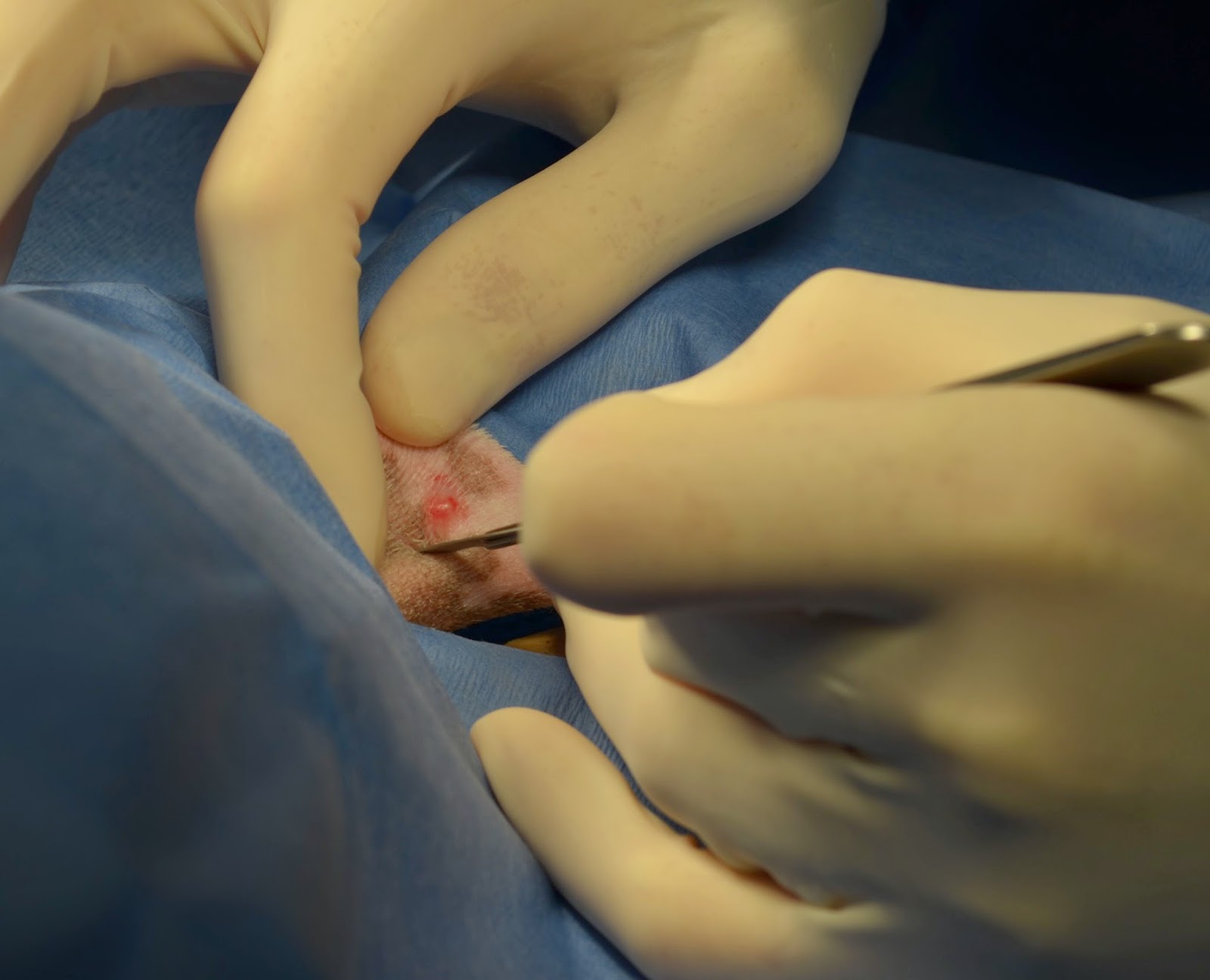 |
| The area around the surgical site was covered with a sterile drape. The incision was made using a scalpel blade (very very sharp!). |
 |
| All sewed up! There is a little bit of blood and serous fluid (liquid of blood that does not contain cells) oozing out. This is totally normal. |
Heidi had the tumors on both back legs removed and her teeth cleaned without any major issues discovered. Everything complete, she was moved to her cage for recovery which will be described in the next post!
Mast cell tumors can be very bad or ok. Heidi's have all been in her skin and removed completely. As of today, she has had 6 removed. She is a mast cell making machine! I keep trying to convince her to stop!
No comments:
Post a Comment