Pages

Sunday, August 24, 2014

SMILE - Post-Anesthesia Recovery

All is done!  The teeth have been cleaned, the veterinarian has performed their examination, and it is finally time to wake up!

The inhaled anesthesia is turned off so that pure oxygen is given for a few minutes.  The monitoring probes and clamps are removed while an assistant prepares the cage with a heating pad and dry bedding.  The oxygen is stopped, the endotracheal tube is disconnected, untied, and the balloon deflated.  Since dental cleanings are wet affairs, the head is dried with towels or a hair dryer.

Your pet is carefully carried over to the cage and placed on the heating pad covered with bedding - usually towels or synthetic lambswool.  

Heidi's recovery cage.  

The endotracheal tube is not removed until your pet is awake enough to swallow on their own.  The risk of removing the trach tube too soon is that the animal will aspirate (inhale) fluid into their lungs.  Fluid in lungs = bad news.  An assistant or LVT will watch constantly to monitor the pet's ability to swallow.  Once the animal is conscious enough to swallow on their own, the trach tube can be removed.  This is why Heidi's tongue in the picture above is pulled out the side of her mouth; when she swallows, her tongue with curl up.  After a few good swallows, the tube is pulled.  Then, time takes over.  A nap and some quiet observation of their surroundings lead to full consciousness and bodily control.

Heidi has been extubated (endotracheal tube removed).
Perfect time for a good, cozy nap.

After your pet has recovered from anesthesia and is comfortably accommodated at home, you may notice a lack of energy and propensity to sleep for the rest of the day and probably the next day too!  This is normal.  When Heidi came home, she could hardly keep her eyes open.  She and the other dogs slept the rest of the day and almost all of the next day.  Usually, your veterinarian will send you home with some home-care instructions.  There may be medications to be given at home, recommendation to feed soft or canned food for a specified period of time, and/or leash walks for a specified period of time.

Heidi can hardly keep her eyes open.

Cuddled under the quilt, she can hardly keep her head up.

Since Heidi had tumors removed from her back legs, she had to visit the vet's office to have her sutures removed after 10-14 days.  Because her teeth were cleaned, Heidi had a few days of canned food since her gums can be sensitive afterwards.

The saga is over - for now!

While dental cleanings can cost a great deal, they are a sure-fire way to maintain and improve the health of your pet.  You may even notice afterwards that they act like a much younger self.

Animals are very good at hiding pain so you may not even know that anything was bothering them until they get their teeth cleaned!

Tuesday, August 12, 2014

SMILE - Surprise Tumor Removal!

(slightly) GRAPHIC PHOTOS IN THIS POST!!  Do not read further if you do not want to see them.


While in the hospital to have Heidi's teeth cleaned, we found two little bumps on her back legs that turned out to be mast cell tumors.  This account is about how we found them, what they are, and how they were removed.


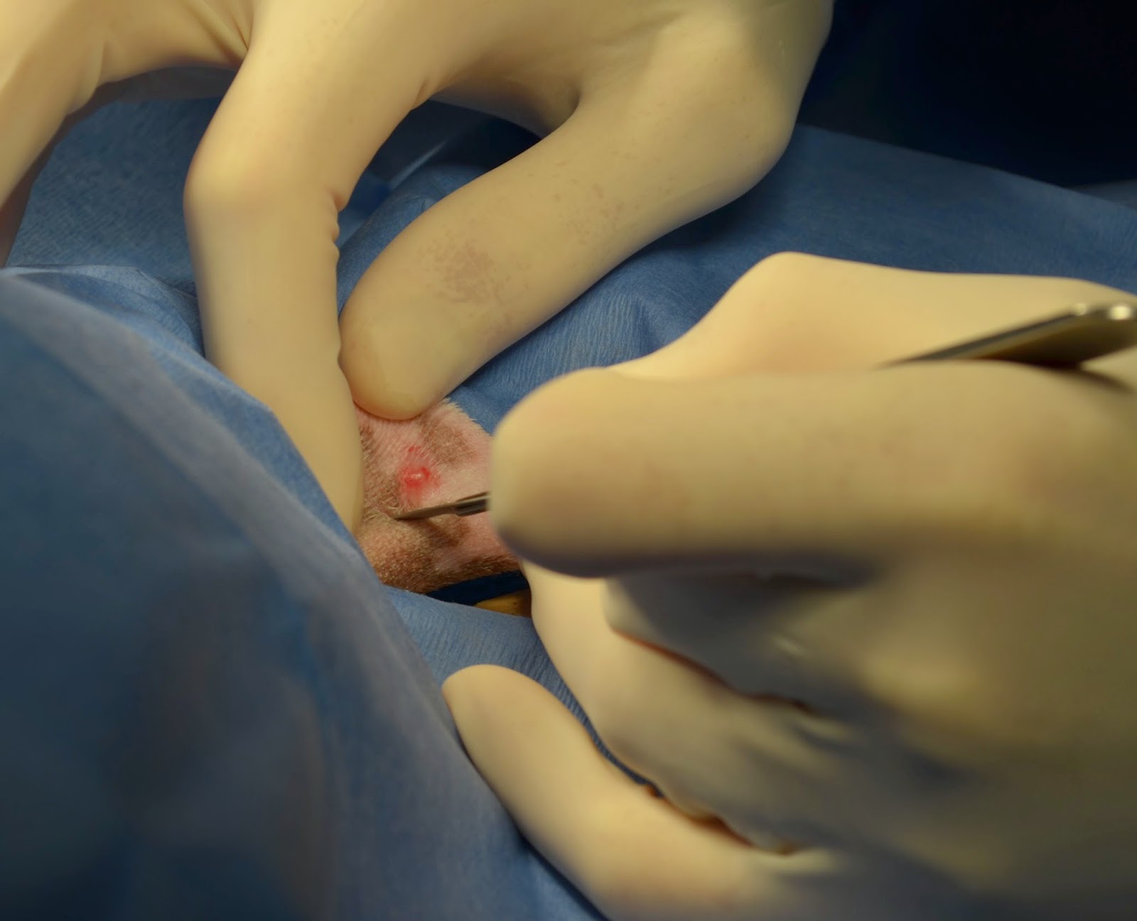
In a previous post, SMILE - Pre-anesthetic Physical and Pre-Medication, I mentioned how the licensed veterinary technician had a difficult time placing Heidi's IV catheter.  Sometimes this can be because the animal is not cooperating - they tend to not understand why we are poking them with sharp objects!  In this case, Heidi's cephalic vein (the one in her front leg) was the one not cooperating.  So, we decided to try to place one in her back leg in the lateral saphenous vein.  When placing an IV catheter, the area is shaved and cleaned to reduce the chance of infection occurring where the catheter enters the skin.  Well, to our surprise, the shaved area revealed a small bump.  It had no hair growing on it and was slightly raised.

The easiest, quickest, and least expensive way to assess a lump is to perform a fine needle aspirate (or biopsy) and cytology.  A thin needle attached to a syringe is inserted into the lump and cells are pulled into the syringe.  The cells are then pushed out of the syringe and onto a glass slide.  The slide, or rather the cells on the slide, are stained and examined under a microscope (cytology).  This LINK has some easy to read information on mast cell tumors, including a picture of what they look like.  Their appearance is distinctive and easy to identify.

Wikipedia's Mastocytoma Picture.
Mastocytoma = mast cell tumor.

Mast cells have a dark blue staining nucleus (center) with dark purple staining granules in the cytoplasm (dots in the area around the nucleus).  

The little lump we found, which was around 3 mm in diameter, showed mast cells in the cytology.  Mast cell tumors in the skin are easily removed when they are small, so we decided to try to place the IV catheter in Heidi's other back leg.  Well, when we shaved the other leg, we found another little mass(!!) in the same place as the first one.  When the vet performed the aspirate and cytology on the second lump, he found more mast cells!  The surgery to remove the tumors was performed before the dental cleaning.  

When an animal undergoes the physiological stress of anesthesia, the time they are anesthetized is minimized as much as possible.  The vet wanted to make sure there was enough time to remove the tumors since that was more important, at that time, than having her teeth cleaned, so the surgery was performed first.  (Luckily, we had enough time to do both!)

Once the IV catheter had successfully been placed, the LVT anesthetized and intubated Heidi in order to start the surgical preparation for the tumor removals.  This is described in the previous post SMILE - General Anesthesia and Monitoring.

The area where the surgery will occur needs to be shaved and cleaned.  Both legs were shaved so that there was a few inches clear around each tumor.  These areas were scrubbed three times alternating with chlorhexidine or iodine scrub (a soapy disinfectant) and alcohol (to clean off the soap and dirt).  Heidi was moved to the surgical suite onto a heated surgery table so she would not get cold.  Anesthesia causes heat loss, as does lying on a cold metal table.  For this reason, heating pads are used to help keep an animal warm during surgery.  Even on the table where dental cleanings take place and in the cage during recovery, there are heating pads.

Heidi's leg has been "prepped" for surgery.
The area has been shaved and cleaned.

The LVT opened the surgical gown and glove outer wrappings as well as the surgical pack and anything else that might be used during the surgery such as a scalpel blade and suture material (the thread used to close the incision).  The vet put on a surgical mask and cap, scrubbed his hands, donned a sterile surgical gown, and carefully put on a pair of sterile surgical gloves. 

Here you can see the surgical instruments, sterile gauze, and suture material.

The area around the surgical site was covered with a sterile drape.
The incision was made using a scalpel blade (very very sharp!).

All sewed up!
There is a little bit of blood and serous fluid
(liquid of blood that does not contain cells) oozing out.
This is totally normal.

Heidi had the tumors on both back legs removed and her teeth cleaned without any major issues discovered.  Everything complete, she was moved to her cage for recovery which will be described in the next post!

Mast cell tumors can be very bad or ok.  Heidi's have all been in her skin and removed completely.  As of today, she has had 6 removed.  She is a mast cell making machine!  I keep trying to convince her to stop!  


Thursday, August 7, 2014

SMILE - Cleaning Teeth

Finally, we are ready to actually clean the teeth!

The licensed veterinary technician performs the cleaning much as a dental hygienist does with people.  The LVT will start by assessing the overall health of the mouth by looking at the teeth to see if any are broken or missing, noting any discoloration, excessive tartar buildup, and/or excessive wear; by looking at the gums to note any redness, swelling, or presence of pus; by looking for the presence of a gingival pocket which is a space where the gums have become detached from the tooth; and/or looking for tumors or gingival hyperplasia (excessive growth of gum tissue); and by looking at the tongue, cheeks, and other structures of the mouth and throat that are visible.

Here you can see Heidi's dirty teeth.  There is a little bit of blood on her upper back teeth.
This happens when your gums are not very healthy.

Dental scaling is the term for cleaning the teeth.  A pointy metal instrument scrapes the tartar from the visible portion of the tooth as well as a short distance under the gumline.  Dental cleanings mainly involved the use of these hand scalers.  However, these days the majority of veterinarians use a dental cavitron scaler which is a small handheld device with a metal tip which sprays water or a solution of chlorhexidine (a disinfectant) and water combined with ultrasonic pulsations.  It's the use of ultrasonic waves that makes cleaning teeth less painful and time consuming and much easier than hand scaling.

The ultrasonic scaler can be seen above.

Scaling the teeth, whether by hand or machine, creates a roughened surface that is a magnet to new tartar growth.  If this roughened surface is not smoothed, it would be worse than not having your teeth cleaned at all!  This is why teeth are polished after they are cleaned.  Keeping tartar away is the key to good dental health.  Tartar builds up on teeth after 24 hours of being dirty.  This is why we, as humans, generally clean our teeth at least twice a day.  This keeps tartar from firmly attaching to our teeth.  This is also why brushing our pet's teeth is a kind way to keep their teeth healthy.  Brushing keeps their mouths healthy which keeps them healthy!

Once the cleaning and polishing is complete, the LVT will get the veterinarian to examine the mouth and share any observations noted during their initial examination.  The veterinarian performs their own examination with a periodontal probe.  The probes have markings on them to show varying lengths in millimeters.  This allows them to measure how deep a gingival pocket is.  The deeper the pocket the more chance there is for bacteria to enter the bloodstream.  This bacteria travelling to the heart is what can cause infections that damage the heart valves and cause other life-threatening complications.

The periodontal probe shown has very faint markings.
The bottom two are 3mm each and the upper two are 2mm each.

Look how far back Heidi's teeth go!

During the examination, the veterinarian uses a chart to keep track of the observations.  Missing or broken teeth, which teeth are extracted, presence of gingival pockets and how deep they are, presence of tumors or hyperplasia, etc. are all noted on the this card which is attached to the chart as part of the permanent medical record.

Mojo's chart is shown (see his feet?)  Circled teeth are missing.
X's are teeth that have just been extracted.

Luckily, Heidi did not need any teeth pulled this time!

Shiny, white(ish), and clean!

Next will be a post about the tumors we found (when the techs tried to place Heidi's IV catheter) and how they were removed.  For not very weak stomachs (only due to mildly graphic pictures)!